
ALARA in Radiology:
Applying Time, Distance and Shielding in Practice
Key takeaways
- ALARA means as low as reasonably achievable. In medical imaging, the goal is to avoid unnecessary radiation while preserving the information needed for diagnosis or treatment. [1,2]
- Time, distance and shielding are practical tools for applying ALARA. Their effectiveness depends on the procedure, equipment, staff position and clinical task. [9-11]
- Occupational dose limits, professional recommendations and facility investigation levels are different things. None should be treated as a routine exposure target. [4,5,7]
- ALARA+ extends the discussion to the physical burden of protection in interventional medicine. It is a professional initiative, not a product certification. [12]
ALARA is easy to recite and harder to apply at the table, mid-procedure, when the clinical priority is in front of you and radiation exposure is invisible. The useful question is not simply, "Can we use less radiation?" It is, "Can we achieve the clinical objective with less unnecessary exposure?" [1,15]
This guide translates ALARA in radiology into practical decisions about imaging technique, working positions, shielding and dose monitoring. It focuses on X-ray imaging, particularly fluoroscopy and image-guided procedures, while distinguishing the protection of staff from the medical exposure of patients.
What ALARA means, and where it comes from
ALARA is the optimization principle within the International Commission on Radiological Protection's framework of justification, optimization and dose limitation. Justification asks whether an exposure is warranted; optimization asks how to carry out a justified activity with appropriate protection; dose limitation restricts occupational and public exposure where applicable. Time, distance and shielding are practical methods, not a substitute for that broader framework. [2,7]
Justification is not a one-time permission to use radiation indefinitely. Each examination or additional imaging step needs an appropriate clinical reason. For patients, optimization means obtaining adequate diagnostic or procedural information with an appropriately selected dose. An image that cannot answer the clinical question is not a successful low-dose examination. [1,15]
Radiological protection uses the linear no-threshold model as a prudent basis for managing low-dose cancer risk. That is a protection assumption, not proof that every very small exposure causes an identifiable injury. It supports avoiding unnecessary exposure without overstating risk or discouraging beneficial imaging. [3]
ALARA also has explicit regulatory applications. NRC licensees, for example, must use procedures and engineering controls to keep occupational and public doses ALARA to the extent practical. Which requirements apply to a particular practice depends on its radiation sources, jurisdiction and regulatory program. [5,6]
Occupational radiation exposure limits: know the source and the quantity
A dose limit is not a dose budget to use up. It also cannot be understood without its jurisdiction, time period and dose quantity. The following are selected reference points, not a complete compliance table.
Source / scope | Selected limit | Important distinction |
OSHA: 29 CFR 1910.1096 | 1.25 rem (12.5 mSv) per calendar quarter | Whole-body category in Table G-18; see (b)(2) and other applicable provisions. [4] |
NRC: 10 CFR 20.1201 | 50 mSv (5 rem) per year | Adult total effective dose equivalent in NRC jurisdiction; additional organ limits apply. [5] |
ICRP recommendation | 20 mSv/year averaged over 5 years | Occupational effective dose, with no more than 50 mSv in a single year. A recommendation, not automatically U.S. law. [7] |
NRC: skin or extremity | 500 mSv (50 rem) per year | Adult shallow-dose equivalent to skin or any extremity; not the whole-body effective-dose limit. [5] |
Selected occupational reference values. Not a substitute for the facility's applicable regulations or radiation-protection program.
The NRC does not regulate the use of X-ray machines; those machines are generally regulated by the states. Do not assume that an NRC number alone establishes the legal requirement for a hospital fluoroscopy service. The facility's radiation safety officer or other designated radiation-protection professional should identify the rules that apply. [6]
Effective dose, equivalent dose to an organ, and shallow dose to skin or an extremity are not interchangeable measures. A higher extremity limit does not mean a worker should accept avoidable hand exposure. Eye-lens, pregnancy-related and other requirements also need separate consideration. [5,7]
Patient medical exposures are not governed by occupational dose limits. Their protection depends on justified imaging and optimization for the clinical purpose. Diagnostic reference levels can help identify protocols that warrant review; they are not individual patient dose limits. [7,15]
Time: reduce unnecessary beam-on time and optimize technique
Dose generally increases with exposure time when other conditions remain unchanged. In fluoroscopy, however, total dose also depends on pulse rate, dose per pulse, acquisition settings, patient size and imaging geometry. Fluoroscopy time alone is therefore not a complete measure of radiation dose. [10,11]
Prepare positioning and equipment before activating the beam. Use last-image hold or stored images when live imaging is unnecessary. Limit cine or other high-dose acquisitions to what the procedure requires, and select the lowest pulse rate and dose mode that provide adequate image quality and temporal resolution for the task. These decisions belong within validated protocols and the operator's clinical judgment. [1,11]
A randomized study of 363 patients undergoing transradial coronary angiography, with or without intervention, illustrates the opportunity. Using fluoroscopy at 7.5 rather than 15 frames per second was associated with approximately 30% lower operator dose and 19% lower patient dose-area product overall, without a significant difference in fluoroscopy time. [8]
Those findings support protocol optimization; they do not promise a 30% reduction in every room or establish that lower rates are suitable for every procedural step. Pulse settings, image quality and clinical needs still have to be evaluated on the system being used. [8,11]
Collimation, patient positioning and appropriate imaging geometry also belong in the discussion. Reducing unnecessary irradiated area and avoiding avoidable repeat imaging can benefit both patient protection and the occupational environment. ALARA is broader than simply spending fewer seconds on the pedal. [1,11]
Distance: use the inverse-square principle without oversimplifying scatter
Intensity varies inversely with the square of distance for an idealized point source with otherwise unchanged conditions. At twice the distance, intensity is one-quarter as great. For example, moving from 1 meter to 2 meters would reduce relative intensity from 100% to 25% in that simplified model. Exposure time and source output must remain unchanged for the same dose comparison. [9]
That calculation explains why distance is useful. It is not a measurement of a staff member's dose in a procedure room. The patient is an extended scatter source, and C-arm angle, shielding, body position and the irradiated area influence the dose at a particular location. The actual reduction from a step back will vary with those conditions.
In practice, increase distance from the patient when the clinical task allows, especially during image acquisitions when close access is not required. Give circulating staff a practical place to work away from the scatter source, and place monitors and equipment so unnecessary proximity is not built into the workflow. [10,11]
Distance should support patient care rather than interfere with it. When staff must remain close, the protection plan needs to account for that working position through appropriate technique, shielding and protective apparel.

Shielding: build protection in layers
During fluoroscopy, the patient is the principal source of scatter reaching staff. The X-ray tube produces the primary beam. Shielding should interrupt relevant radiation paths without interfering with imaging, sterile access or patient care. Neither the word "lead" nor a large lead-equivalence number makes placement unimportant. [10]
Structural shielding protects designated occupied areas, such as control positions and adjacent spaces, according to the room's design. It does not automatically protect personnel standing inside the procedure room beside the patient.
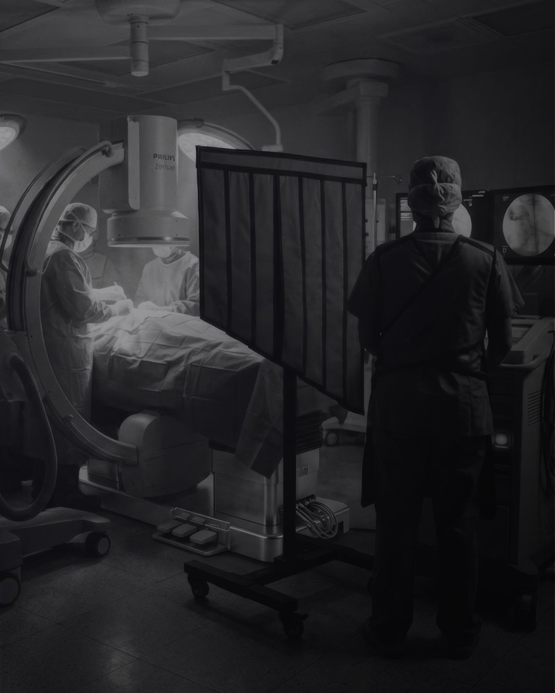
Equipment-mounted shielding, including ceiling-suspended screens and table-side curtains, helps protect personnel working near the table. Gaps and working positions matter, and shielding should be reassessed when the C-arm, table or staff position changes. Positioning shielding between the patient, the main source of scattered radiation, and staff intercepts that scatter before it reaches the protected zone.
Mobile barriers can provide additional protected working positions for staff whose tasks permit them to remain behind the shield. Personnel outside its coverage do not receive the same benefit.
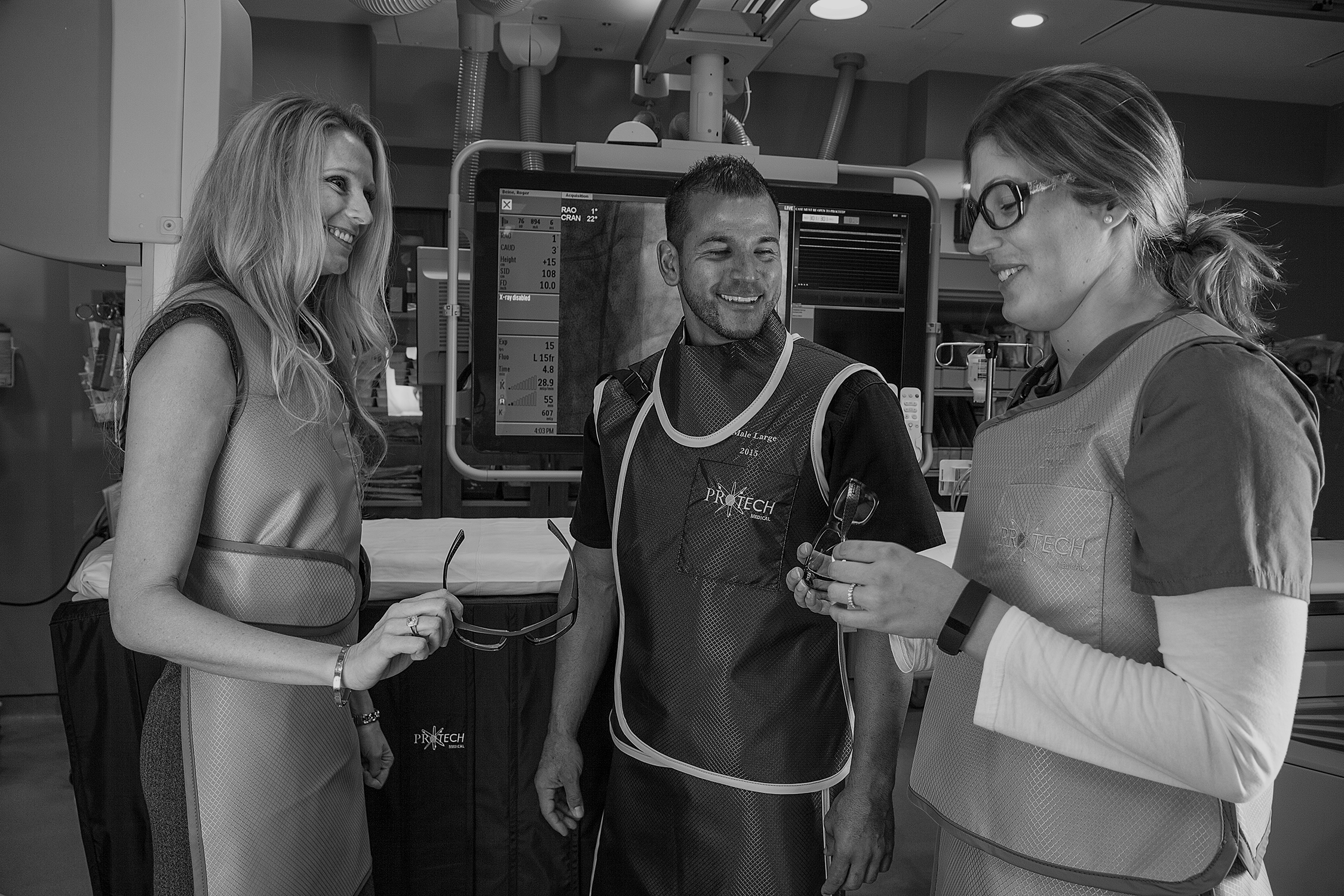
Personal protective equipment protects the portions of the wearer covered by the device. Aprons, thyroid collars and radiation-protective eyewear should be selected for the exposure conditions and facility program, including fit, lateral coverage and movement during the procedure. [10]
Radiation-attenuating gloves require particular care. They are not permission to place hands in the primary beam. If an attenuating glove enters the fluoroscope's exposure-control sensing region, the system may increase output. Hand protection should be addressed through positioning, technique and task-specific guidance, not a routine glove recommendation for everyone. [10]
Layered protection does not create one universal reduction percentage. A result measured at an operator's eye in one shielding configuration cannot automatically describe whole-body exposure, another staff position, or a different manufacturer's equipment. Compare products using relevant test conditions and assess performance in the actual working environment.
Monitoring: turn ALARA into a reviewable process
A personal dosimeter measures exposure; it does not shield the wearer. Wear the assigned monitors in the locations specified by the radiation-protection program and return them on schedule.
For image-guided interventional work, IAEA guidance recommends two dosimeters: one outside the apron near the collar and another under the apron on the torso. Other roles or exposure conditions may require a different arrangement. Confirm the correct arrangement with the radiation-protection team before changing badge placement. [10]
Review unusual readings rather than waiting for a legal limit to be approached. Workload, procedural complexity, monitoring practices and equipment changes can help explain a trend. Local investigation levels should trigger appropriate review; they are not interchangeable with statutory limits. [5,10]
For patients, review the modality-appropriate dose indicators alongside image quality and clinical results. The purpose is to identify avoidable exposure and improve practice, not to compare unlike examinations solely by their numerical dose values. [1,15]
ALARA+ in 2026: addressing radiation and physical burden
In March 2026, nine medical societies called for ALARA+: As Low and As Light as Reasonably Achievable in interventional medicine. The initiative addresses occupational radiation exposure alongside the orthopedic burden of protective equipment and the working environment. [12]
ALARA+ is not a repeal of conventional ALARA, a new statutory dose limit, or a certification attached to an apron. It also is not simply an instruction to buy a lighter garment. Engineering controls, enhanced shielding, ergonomics and team-wide protection are central to the discussion. [12]
For apparel selection, evaluate weight alongside shielding performance, fit, coverage and how the load is carried. A lighter core or a different garment configuration may be useful, but the complete arrangement still needs to meet the facility's protection requirements. Compare documented garment performance and fit rather than relying on a material name alone. [10,12]
How Protech supports ALARA in medical imaging
Protech manufactures custom Proguard radiation-protective lead aprons in South Florida and offers additional product categories including leaded eyewear, thyroid protection, radiation-attenuating gloves, barriers and drapes. These products can support an ALARA program when selected and used for the appropriate application. They do not, by themselves, make a procedure or facility compliant. [13]
For teams evaluating garment weight, Nano and Premier are among Protech's core-material options. Compare the specific formulation, garment configuration, coverage and documented attenuation at relevant radiation qualities. Proguard core material options have different specifications, and total garment weight also depends on the style, size and coverage selected. [14]
Selection starts with the actual task: who is exposed, where they work, what movement is required and which shielding is already available. Protech can help compare product configurations; the facility's clinical and radiation-protection teams determine how those products fit the approved protection program.
Keep ALARA in the daily workflow
Before imaging, confirm the clinical purpose, select the appropriate protocol and identify the staff positions that need protection. Check the condition and placement of required shielding and confirm that monitoring arrangements are understood.
During the procedure, use live imaging only when needed, maintain clinically appropriate technique and reassess coverage when geometry changes. Afterward, review relevant dose information and investigate unexpected findings or equipment concerns through the facility's established process. [1,5,15]
The strongest ALARA program is not defined by the lowest number on one case or the highest rating on one product. It is a repeatable process that supports useful imaging, appropriate staff protection and better decisions over time.
To compare radiation-protection options for your workflow, contact Protech.

Frequently asked questions
What does ALARA stand for in radiology?
ALARA stands for as low as reasonably achievable. In X-ray imaging, it means avoiding unnecessary exposure while retaining the image quality and information needed for diagnosis or intervention. It does not mean minimizing dose without regard to clinical benefit. [1,2]
What are the three principles of ALARA?
Time, distance and shielding are commonly taught as three practical methods for reducing external radiation exposure. The broader ICRP framework is justification, optimization and dose limitation. ALARA is the optimization component of that framework. [2,9,10]
What is the occupational radiation dose limit in the United States?
There is no single number that describes every applicable rule. NRC adult total effective dose equivalent is limited to 50 mSv per year in its jurisdiction; OSHA's older Table G-18 uses a quarterly whole-body framework. State X-ray rules and additional dose limits must also be considered. Confirm requirements with the facility's radiation-protection lead. [4-6]
Does doubling distance always reduce staff dose by 75%?
That is the idealized inverse-square result for a point source under unchanged conditions. Actual patient-scatter exposure depends on source extent, geometry, shielding and staff position. Use distance where practical without treating one step back as a guaranteed reduction. [9,10]
Is an apron "ALARA certified"?
ALARA is an optimization principle and program obligation where applicable, not a stand-alone product certification. Evaluate an apron using its documented performance, intended use and the facility's requirements. ALARA likewise does not endorse a specific Protech material or model. [2,5,12]

Let's talk Protech-tion |
Or call 561.627.9769 to get started.
References
Sources checked September 15, 2026. Numbered citations in the article correspond to these linked references. Regulatory and manufacturer information should be rechecked before publication if the draft is held for an extended period.
1. FDA. Medical X-ray Imaging.
3. ICRP Publication 99. Low-dose Extrapolation of Radiation-related Cancer Risk.
4. OSHA. Ionizing Radiation: Standards. See 29 CFR 1910.1096(b), Table G-18, and applicable provisions.
5. NRC. 10 CFR Part 20. See sections 20.1101, 20.1201 and 20.1502.
6. NRC. Agreement State Non-Emergency Concerns. Identifies state responsibility for X-ray machines.
9. OSHA. Ionizing Radiation: Ionizing Radiation Equations. Inverse-square relationship.
13. Protech Medical. Proguard Lead Aprons. Manufacturer product and manufacturing-location information.